|
|||||
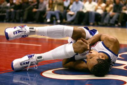
Partial Thickness Defects The term “partial thickness defects” indicates damage of the zonal articular cartilage, without penetration into the underlying bone. This type of injury on the surface leaves the defect site inaccessible to the cell types that stimulate self-healing responses. The macromolecules in articular cartilage (including proteoglycans) make the tissue surfaces anti-adhesive. While chondrocytes begin to synthesize extracellular matrix (ECM) immediately after the injury, not enough chondrocytes are able to migrate to the affected sites for full regeneration to occur. Self-healing activity gradually comes to a stop, resulting in progressive reduction of tissue function, initiating tissue degeneration. |
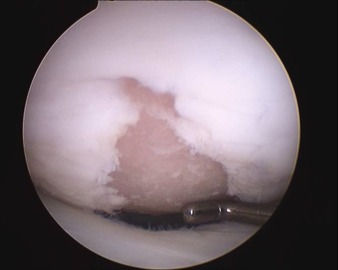
Full Thickness Defects The more severe case of cartilage defect is full thickness
defect, penetrating into the bone, past the calcified zone (a layer of
calcified cartilage covers the underlying bone). Unlike partial thickness
defects, the site is accessible to blood cells, bone cells, and progenitor
cells in bone morrow, all of which are involved in a spontaneous healing
process. Upon injury, the defect site is immediately filled with a fibrin clot
and an inflammatory response is activated. Mesenchymal stem stems located in
the bone marrow move into the defect site, replacing the fibrin clot completely
after approximately one week. These stem cells then differentiate into
chondrocytes, due to the chondrocytes’ ability to secrete proteoglycan-rich
extracellular matrix, helping repair the damaged cartilage tissue. However, healing is short-lived as the new tissue is of inferior quality. |
||||