The image below illustrates the zonal arrangement of articular cartilage. 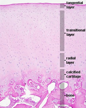 Middle (Transitional) Zone The middle zone serves as a transition 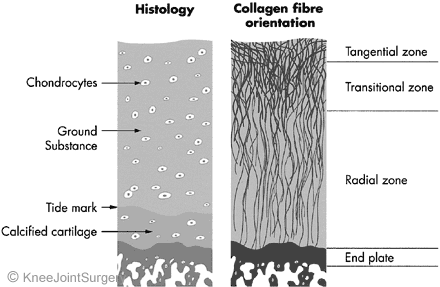 |
Articular cartilage is a heavily hydrated tissue with 75% of the tissue's weight being water. The solid matrix consists of about 75% of collagen and 25% proteoglycans. The principal collagen type in articular cartilage is type II collagen. Proteoglycans are large sugar molecules that are negatively charged and, therefore, retain a large portion of the tissue's water. This soft, hydrated tissue exhibits remarkable biomechanical properties. Though articular cartilage is not very stiff, it is very strong in compression and it is remarkably durable. Despite the fact that cartilage cells are very few, very slow, and lack nutrients (and so they do not turn the tissue over), articular cartilage operates as an almost frictionless surface for many decades of life. When one looks at a side view of cartilage, one can identify four distinct zones: superficial (or tangential), middle (or transitional), deep (or radial), and calcified. Each zone has its unique matrix composition, morphology, cellular, mechanical, and metabolic properties. Superficial (Tangential) Zone The superficial zone accounts for approximately 15% of the total thickness; however, it contains the highest collagen density. Collagen, a group of naturally occurring proteins, is abundant in cartilage in the form of fibrils. In this zone, the fibrils are thin, densely packed, and form an oriented lamina covering the joint. Fibroblast-like chondrocytes with few organelles lie along the superficial zone, oriented parallel to the surface, in the direction of sliding where shear stress develops. The way the components of this zone are organized contributes to its shear resistance and tensile strength.  |
|||||
|
||||||