Microfracture Surgery
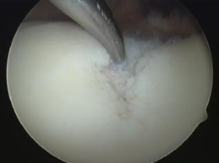
Microfracture surgery is the most common method used to treat smaller articular cartilage defects. The surgery is performed by arthroscopy. Once the joint has been cleaned of calcified cartilage, the underlying subchondral zone is drilled and shaved to create small fractures. This triggers the release of bone marrow progenitor cells forming a blood clot, similar to the self-healing response that occurs in full-thickness defects. The blood clot releases cartilage-building cells. While this treatment is effective for small defects (<2 cm sq.) and attractive due to low morbidity, minimal invasion, short surgery time and short recovery time, it has faults. Sometimes, little or no hyaline cartilage is regenerated. The remaining cartilage will also turn into weaker fibrocartilage, providing less mobility.
In microfracture surgery, using an awl, fractures are made in the underlying subchondral bone, as pictured above, to trigger a self-healing response.
|
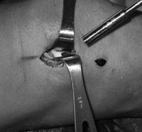
Autografts and Allografts For an autograft, healthy, cylindrical cartilage tissue plugs are harvested from the low weight-bearing area of the knee and then implanted into defect sites, similar to an autologous chondrocyte implantation. However the lack of tissue donors, the graft instability, and surface mismatch of the graft are all issues that arise. In addition, considering that the tissue was harvested from a low weight-bearing area, it may have difficulty surviving in a high weight-bearing area. This technique is best for smaller size defects (1-4 cm sq.).
Shown above is a small cylindrical defect on one of the condyles of the femur of the knee. Allografts use cartilage tissues from tissue banks, alleviating the insufficient donor tissue supply. They also require multiple surgeries similar to autografts. The limitations are similar, including contour matching and load-bearing capacity. This method may also cause immune reactions, such as inflammation or infection. Allografts contain dead cells that can be problematic due to their inability to maintain the articular surface. Autologous Chondrocyte Implantation
Autologous chondrocyte implantation is a two-step surgery using cell transplantation techniques for cartilage repair. This treatment is recommended for patients with cartilage lesions between 1 cm sq. and 12 cm sq. thick, or for those who have undergone previously failed microfracture surgeries. In the first surgery, a small piece of healthy cartilage is removed from the lower area of the knee. Chondrocytes are retrieved from the tissue and expanded in vitro for 3-5 weeks. The defect site is then trimmed and prepared for the second surgery in which the grown cells are injected into the site. The chondrocytes are then sealed into the defect site with a periosteal patch taken from the shin bone. The periosteum is the thick tissue that covers the shin bone. This process has limitations including the long recovery time and the need for multiple surgeries. Also, de-differentiation of the patient’s cells during the period of cell culturing in vitro, and decreased human chondrocyte numbers due to aging may impair the use of autologous chondrocyte implantation. |